White Shark Bite Forensics -
a Photo Essay
 |
A large White Shark obligingly chomps down on our seal-shaped decoy, leaving a terrific set of dental impressions in the hard plastic coating that penetrate deep into the compressed foam interior. Rubbery on the outside, sort of like sponge toffee on the inside. Yum! |
Here, Rick Allen of Nautilus Productions examines the shark-bitten decoy. Ever the consummate professional, the first thing he checks is how his 'lipstick camera' tolerated the abuse of being chomped by a "big 'ol" White Shark. He doesn't realize it yet, but Rick is about to receive a 'crash course' in shark tooth terminology from yours truly. |
 |
|
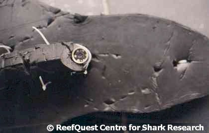 Overview of the undersurface of the decoy, showing the 'lipstick
cam' in situ. Notice that many of the tie wraps (a.k.a.
'zap straps') that fasten the camera to the decoy's keel have been
severed. The camera survived its ordeal just fine - it was the
wimpy connector cables that gave out.
Overview of the undersurface of the decoy, showing the 'lipstick
cam' in situ. Notice that many of the tie wraps (a.k.a.
'zap straps') that fasten the camera to the decoy's keel have been
severed. The camera survived its ordeal just fine - it was the
wimpy connector cables that gave out.
A sketch of the overall decoy, showing the upper tooth impressions left by an earlier White Shark attack. The bite diameter is 10.4 inches (26.5 centimetres) across at the level of the second lateral teeth (L2). The maximum 'reach' of the bite is 6.9 inches (17.4 centimetres).
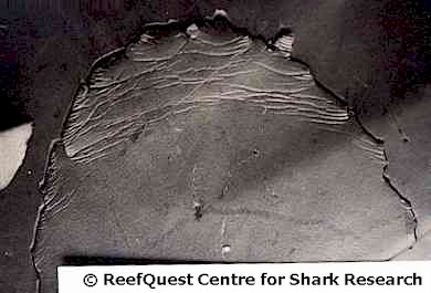 Shot of the undersurface of the board, showing the impressions left
by the upper jaw teeth of a large White Shark. Notice how broad,
flattened and regularly spaced the tooth punctures are - this is what
gives them away as upper tooth punctures.
Shot of the undersurface of the board, showing the impressions left
by the upper jaw teeth of a large White Shark. Notice how broad,
flattened and regularly spaced the tooth punctures are - this is what
gives them away as upper tooth punctures.
Also note that the second impression to the right of the mid-line of the arc has a strangely 'crinkled' flap attached to it - quite different from the blade-like stabs that characterize the other tooth impressions. When the decoy was initially recovered by deck hand Alison Kock, we believed this puncture is the one that had a White Shark tooth embedded firmly in it. In normal predation (on somewhat more digestible objects), the upper jaw teeth act as knives, to saw out bite-sized hunks of flesh.
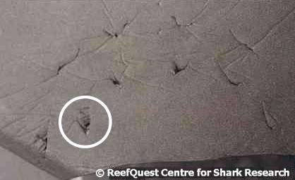 Shot of the upper surface of the board, showing two distinct series
of arcs caused by the lower teeth of the attacking White Shark.
Notice that these punctures are less regular in spacing and
distribution than those caused by the upper teeth, as well as less
broad and less flattened - this is what marks them as lower tooth
impressions. Notice that the second tooth impression from the
left edge (circled) also has a crinkled flap. This is an important clue
that our embedded tooth may not have been an upper tooth, as we first
thought.
Shot of the upper surface of the board, showing two distinct series
of arcs caused by the lower teeth of the attacking White Shark.
Notice that these punctures are less regular in spacing and
distribution than those caused by the upper teeth, as well as less
broad and less flattened - this is what marks them as lower tooth
impressions. Notice that the second tooth impression from the
left edge (circled) also has a crinkled flap. This is an important clue
that our embedded tooth may not have been an upper tooth, as we first
thought.
The lower jaw teeth of White Sharks are less flattened and blade-like than the uppers because they serve different functions. In normal predation, the lower teeth act as the prongs of a fork, holding the food item securely while the uppers carve out a manageable piece.
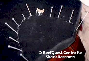 Photo
showing the undersurface of the decoy with the embedded tooth re-inserted in
the same puncture from whence we originally believed it came. The white objects bristling
from the puncture wounds are tie wraps. I inserted one of these at the
deepest point in each puncture to measure its depth and distance from
neighboring punctures. It also helped reveal the bite rather
dramatically.
Photo
showing the undersurface of the decoy with the embedded tooth re-inserted in
the same puncture from whence we originally believed it came. The white objects bristling
from the puncture wounds are tie wraps. I inserted one of these at the
deepest point in each puncture to measure its depth and distance from
neighboring punctures. It also helped reveal the bite rather
dramatically.
Rick Allen came up with the idea of using multiple tie wraps to measure all tooth punctures simultaneously. In his his honor, we jokingly named this technique the Allen Dental Depth Index or ADDI (which, it just so happens, is a documentary film-maker's award - so the acronym has a double significance for its inventor).
Notice that there is an 'extra' puncture, immediately to the left of the embedded tooth. Did the shark re-purchase its upper jaws "to get a better grip", or is this due to the loose embedded tooth 'wiggling' somewhat before it was lost? That the surrounding teeth do not show similar multiple punctures leads me to suspect the latter.
|
|

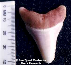
Front (left) and rear (right) views of the embedded tooth. Notice that the serrations on the edges of the tooth blade are coarse and irregular. This characteristic helps to distinguish White Shark teeth from those of certain whaler sharks of the family Carcharhinidae that also have broadly triangular, serrated upper teeth. Note also that the front or "labial" face of the tooth blade is flattened and the rear or "lingual" face is convex (rounded outward). Note also the large cluster of nutritive pores near the center of the tooth base - this is where the living tooth is supplied with blood via tiny blood vessels. If it seems counter-intuitive that the inner face of a shark tooth is convex, recall that the teeth are generated in a membrane inside the jaw in an upside-down position, rotating forward as they develop into their erect, functional position. Because shark teeth develop in an inverted orientation, it is the lingual faces that conforms to the curvature of the front margin jaw when initially formed and become the 'rear' of the teeth when they rotate into their 'upright' functional orientation. I often wonder how many people who wear shark tooth jewelry realize that, by wearing the teeth curved face outward, they are actually displaying the back of the tooth. The arched root shape, the graceful widening of the blade where it joins the root, and the overall roundedness of the lingual face all indicate that this is a tooth from the front of a White Shark's lower jaw.
The vertical height of this tooth is 1.34 inches (3.4 centimetres), its slant height - measured along the longest edge of the tooth from the outer edge of the root to the apex of the blade - is 1.56 inches (3.95 centimetres), and its width across the base is 1.14 inches (2.9 centimetres). The enamel height is 1.04 inches (2.65 centimetres), which can be used to estimate the total length of its former owner. But - as you can see in the photos above - the tip has been broken off via a type of impact-caused structural damage called a "spall fracture". Since no tooth tip was found embedded in the decoy and its composite material simply isn't hard enough to have caused this kind of damage, the fracture probably occurred before the shark obligingly bit our decoy. Fish bones are generally rather delicate, so the tooth tip probably shattered as it struck the much more solid bone of a marine mammal (such as a sea lion or a dolphin, or possibly even a whale carcass). Adjusting for the lost tooth tip and taking into account the diameter of the bite impression in the decoy, I estimate that the attacking shark was about 11 feet (3.4 metres) in length.
Even if we did not know the exact position of the tooth by its location among the upper jaw tooth puncture marks, it is a fairly straight-forward matter to identify the position of origin of an isolated White Shark tooth. My colleague Ralph Collier of the Shark Research Committee - who has studied White Shark teeth and tooth impressions in intimate detail for several decades - kindly and generously taught me some of the fundamentals.
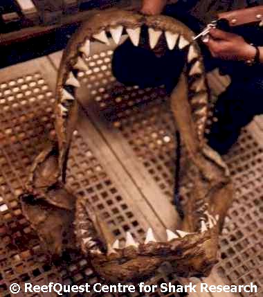 Consider the White Shark jaws at left. These
are the infamous "Port Fairy" jaws at the British Museum of
Natural History (formerly thought to be from a 36.5-foot [11.1-metre]
specimen, a figure which is now known to be erroneous), digitally altered to
replace a missing and a broken upper tooth. Notice that, in White
Sharks, the upper jaw teeth are strongly differentiated by size and
shape. Starting from the symphasis (center) of the jaw and proceeding
toward the jaw corners, there are two almost symmetrical "anteriors"
(Curiously, in White Sharks, it is the second -
rather than the first - upper anterior that is the largest tooth.),
a smaller tooth with a sinusoidal (shaped like a 'stretched-out' S)
cross-section and a reversed orientation (pointing toward the mid-line of
the jaw rather than the jaw corners) called an "intermediate"*,
then four or five larger teeth that are increasingly slanted toward the jaw
corners called "laterals", followed by five or six much
smaller "posteriors". With the exception of those of
the 'backward-mounted' intermediate teeth, the longest cutting edge in White
Shark tooth blades occurs on the side facing the jaw corners. Also, as
an additional clue, the more gracile ('slender') of the two root lobes is
oriented toward the symphasis of the jaw.
Consider the White Shark jaws at left. These
are the infamous "Port Fairy" jaws at the British Museum of
Natural History (formerly thought to be from a 36.5-foot [11.1-metre]
specimen, a figure which is now known to be erroneous), digitally altered to
replace a missing and a broken upper tooth. Notice that, in White
Sharks, the upper jaw teeth are strongly differentiated by size and
shape. Starting from the symphasis (center) of the jaw and proceeding
toward the jaw corners, there are two almost symmetrical "anteriors"
(Curiously, in White Sharks, it is the second -
rather than the first - upper anterior that is the largest tooth.),
a smaller tooth with a sinusoidal (shaped like a 'stretched-out' S)
cross-section and a reversed orientation (pointing toward the mid-line of
the jaw rather than the jaw corners) called an "intermediate"*,
then four or five larger teeth that are increasingly slanted toward the jaw
corners called "laterals", followed by five or six much
smaller "posteriors". With the exception of those of
the 'backward-mounted' intermediate teeth, the longest cutting edge in White
Shark tooth blades occurs on the side facing the jaw corners. Also, as
an additional clue, the more gracile ('slender') of the two root lobes is
oriented toward the symphasis of the jaw.
Since the flattened surface of the tooth blade is the labial or outer surface of the tooth, one can quickly determine that the embedded tooth came from the right side of the lower jaw (the photo of the rear of the embedded tooth shows us how the tooth would be oriented looking forward from inside the shark's jaws - and it is the shark's left or right side that matters here, not our own left or right as we face the shark). The near-symmetry of the embedded tooth tells us that its position in the jaw must have been close to the symphysis, since the blades of White Shark teeth become increasingly 'slanted' toward the jaw corners the farther they develop from the symphysis. To put this another way, the embedded tooth is far too symmetrical to be a lateral or a posterior and lacks the sinusoidal cross-section that characterizes an intermediate tooth. Since the embedded tooth is not quite symmetrical enough to be a first anterior, it must - by process of elimination - be a second anterior. Thus, the embedded tooth removed from our seal decoy was almost certainly an lower, second anterior tooth from an approximately 11-foot (3.4-metre) -long White Shark.
 Having
examined the bite damage to our seal decoy and the embedded tooth from the
stable platform of our hotel room, it was time to return to sea to try our
luck again.
Having
examined the bite damage to our seal decoy and the embedded tooth from the
stable platform of our hotel room, it was time to return to sea to try our
luck again.
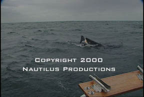
As you can see at right, we were successful in tricking another White Shark into chomping on our decoy. Although this animal didn't leave behind an embedded tooth, from the diameter of the bite I was able to estimate its length at just under 15 feet (4.5 metres).
This is the individual shark that severed Rick Allen's connector cables, rendering his lipstick cam useless. Fortunately, we had already obtained some incredible underwater footage using this technology, so Rick regarded the loss of the connector cables as par for the course. When Rick joins me in the field next year, he plans to bring at least three lipstick cameras. The well-chomped decoy, however, was retired and now rests, unmolested, among my most treasured memorabilia.
* = There is currently some controversy about the use of the term "intermediate" to describe the reduced third tooth on either side of the upper jaw of the White Sharks and most other lamnoids. For the details, Click Here. To return to the Port Fairy jaws text, Click Here.
