Skeleton in the Corset
Of course, teeth are impotent without firm jaws to wield them, backed up by a strong spine and other skeletal supports. The internal skeleton of a shark is composed of primarily of cartilage and connective tissue. It provides firm attachment sites for muscles and prevents the body from buckling under their collective pull. Cartilage is tough and durable, yet relatively light-weight (only about half the density of bone), reducing substantially the amount of swimming effort a shark must invest to prevent sinking.
In certain locations where strength is particularly important - such as the jaws and parts of the backbone - shark cartilage is fortified with tiny, hexagonal crystals of calcium salts called "tesserae" (after the little tiles that compose mosaics). Tesserae give shark cartilage much of the strength of bone without most of the accompanying weight. As a shark grows, however, the mass of its skeleton increases much more rapidly than does its surface area (mass increases as a cube of dimension, area as a square). Therefore, the skeletons of larger animals require proportionally greater strength than do those of smaller ones - which is why, for example, an elephant's femur (thigh bone) is not simply a scaled-up version of a mouse's; rather, it is stouter and with a much larger cross-sectional area. Since a shark's body is supported by water, it does not need thicker limbs as it grows. But one would expect that the jaws and backbones of large shark species would require relatively more strengthening than those of smaller individuals. In a 1991 paper, shark biologist Guido Dingerkus and his co-workers examined the tesserae of over 50 species of modern sharks. They found that the jaws of large specimens of most species had only a single layer of tesserae, but that three species - the Bull (Carcharhinus leucas), the Tiger (Galeocerdo cuvier), and the White Shark - had two, three or more layers depending on overall body size. The greatest number of tesserae layers Dingerkus et alii found in any shark was five, in a 16- and an 18-foot (5 and 5.5-metre) Great White. The White Shark thus has exceptionally heavily fortified jaws, able to withstand the titanic forces to which they are subjected.
The remainder of the White Shark's skeleton is no less elegant. As in other sharks, the skeleton of the Great White can be divided into two basic parts. One of these parts consists of the fins' supporting structures. The supporting structures of the paired fins include transverse "girdles", which firmly anchor the pectoral and pelvic fins in place. The pelvic girdle is relatively simple, little more than a pair of cartilaginous bars fused at the mid-line of the body. But the pectoral girdle is a bit more involved, a six-part U-shaped structure which provides solid attachment points for the pectoral fin skeleton. For all their various functions, most shark fins are built on a single, simple plan. Beginning closest to the body and working outward, this plan features: relatively large blocks of cartilage at the base of the fins; smaller, more finely divided cartilages which seem to fan-out from the basal cartilages (in males, certain basal elements are extended posteriorly, to provide support for the intromittant organs called "claspers"); and, lastly, thin, unsegmented rods of cartilage that extend the fanning-out of the radials almost to the very edges of the fin.
The caudal fin of the White Shark appears nearly symmetrical from the outside, with both lobes similar in size and shape. But underneath, the Great White's caudal fin structure is just as top-heavy as that of other sharks. Unlike the tail of teleost fishes, in which the backbone terminates at the caudal fin base, the backbone of sharks extends almost to the tip of the upper caudal lobe. If the lower lobe were not somehow fortified to provide similar rigidity, the lamnids' characteristic body form and stiff-bodied swimming style would be far less efficient than they are. In the same 1986 paper in which Wolf-Ernst Reif and David Weishampel studied the denticle crown alignment in the Shortfin Mako, they also studied the anatomy of its lunate caudal fin and supporting keels. Reif and Weishampel found that in the Shortfin Mako the serial cartilaginous arches that form a tube through which the body's major bood vessels pass have expanded processes that allow caudal fin muscles and tendons to attach in a complex manner that strengthens both the upper and lower lobe. Further, they found that the armor-like skin covering the keels is 5 to 16 times thicker than that covering the back, further supporting the caudal fin. Both these adaptations enable the Shortfin Mako's upper and lower caudal lobes to function as a single propulsive structure. An opportunistic 'peel dissection' I performed on the caudal fin of a small (5-foot or 1.5-metre) White Shark has revealed that its arch, muscle and tendon structures are essentially the same as in the Shortfin Mako. Measurements by fluid dynamics scientists have demonstrated that such a stiff, oscillating tail is twice as efficient at pushing back water as is a rotating boat propeller. Therefore, much of the Great White's propulsive power is due to details its caudal fin structure.
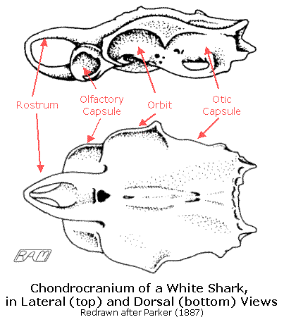
The other part of the shark skeleton consists of the skull and vertebral column as well as associated structures such as the gill bars, jaws and supporting arches. A shark's skull is a relatively simple structure: a one-piece box of cartilage that encases the brain, lacking any trace of the meandering sutures characteristic of the skulls of teleosts and tetrapods (including humans) or the mindbogglingly-complex system of catches and levers so characteristic of teleost skulls. When viewed from above, the White Shark's skull is shaped like a stone arrow-head, with (proceeding from front to back): a tripodal rostrum (snout), followed by globular olfactory capsules that house the scent organs, large, forward-oriented orbits (eye sockets), and a long rectangular otic capsule housing the inner ear mechanism. The backbone or spinal column consists of segmental vertebrae (singular: vertebra), each relatively simple: a centrum composed of two cones fused tip-to-tip (through which the notochord runs), above which is an arch (through which the spinal chord passes) and - in the tail stalk only - beneath which is another arch (through which the body's main blood vessels course). The number of vertebrae a shark has varies considerably, both among and within species. In the White Shark, the total number of vertebrae ranges from 170 to 187. Sharks have no ribs to speak of, but between successive arches are cartilaginous plates which strengthen the spinal column as a whole. The backbone resists the powerful compressional forces induced by the swimming muscles, which would otherwise cause a shark's body to shorten worm-like rather than bend, greatly reducing its swimming efficiency.
|
|
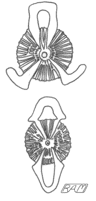
Cross Sections of a White Shark's Vertebrae. The top diagram is of a thoracic vertebra, characterized by having a neural arch (through which the spinal chord extends) only. The lower diagram is a caudal vertebra, characterized by both a neural arch and a haemal arch (through which the dorsal aorta extends). Redrawn from Parker (1887) |
The shark gill bars form a complex 'basket' that supports a shark's delicate gills. The gill bars of the White Shark are composed of a series five pairs of five cartilaginous elements. Together, they form a structure reminiscent of a pair of clawed hands that have been fused at the wrists. The space between successive gill bars is partially bridged by cartilaginous rays that branch from either side like the teeth of a double-sided comb. Those branches on the inside help prevent food from escaping out the gill slits; those on the outside support the feathery gill membranes, the site of gas exchange for the animal. Beneath the skull and anterior to the gill bars are located the jaws of a shark. Shark jaws are unusual among vertebrates in that the upper jaw is not fixed to the skull, a feature which has greatly enhanced the feeding success of the group (about which more in a moment). The jaws are composed of two upper halves - collectively termed a "palatoquadrate" - and two lower halves that form the lower jaw. The jaws are buttressed from behind on each side by a two-part "hyoid arch".
There can be little question that the jaws of sharks in general and the Great White in particular are fearsomely efficient. The jaw suspension of sharks varies considerably from species to species. In the White Shark, the upper and lower jaws articulate well behind the eyes and the palatoquadrate is rather loosely attached to the undersurface of the skull. Because the White Shark's upper jaws are slung loosely beneath the skull rather than fused in place (as in humans), they can be protruded from the head. This enables the Great White to bite prey or objects from almost any angle, despite the underslung arrangement of its jaws. How the White Shark can protrude its jaws far out of the head and yet manage a powerful bite is another astonishing piece of bioengineering. But to understand how this gnathic feat is accomplished, we must first understand something about the muscular system of sharks.
